
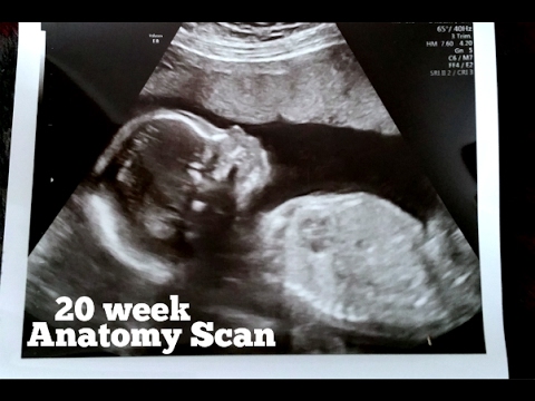
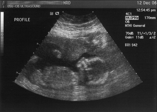
Our 20 week Ultrasound Anatomy Scan. This ultrasound anatomy scan was done on September 21, 2016. At the time I was 20 weeks and 3 days pregnant. My son was born on January 18, 2017 and I can’t wait to post …


More Information About Your Ultrasound Anatomy Scan
An ultrasound scan might appear like a remarkable home entertainment to you. Ultrasound scans can in some cases discover issues with the child, and that’s why they are used. So , you must permit the medical personnel (sonographer or physician) to perform their work, without distracting them.
Be patient! An anatomy scan takes a lot longer than any other kind of pregnancy scan.
If the child is ideally placed by lying in the appropriate position, the scan generally lasts 40 minutes. However, it can take longer.
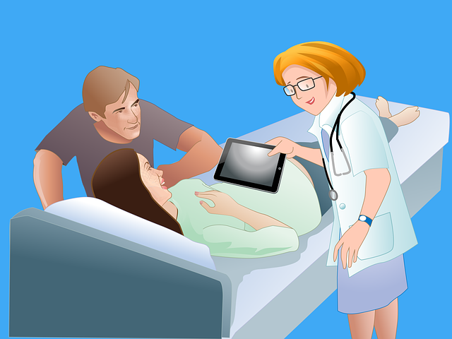 You might be asked to gently move your belly or take a brief walk to get junior to be more visible in the scanner. Throughout the treatment gel is smeared to the stomach location. This is to to guarantee excellent contact in between the skin and transducer (the gadget that produces ultrasonic waves).
How the Scanner is Moved to Obtain the Best Images of Your Baby
The transducer is put on the tummy and is rolled back and forth. Meanwhile, internal images can be seen on a display beside the service technician. In early scans the sonographer (professional) just takes 8 or 12 images, in later scans 40 or 50 might be taken.
Your sonographer or physician may describe the information seen during your ultrasound scan to you throughout or directly after your scan. In some cases, they’ll send out the outcomes to your midwife or medical professional. When that occurs they’ll normally go through things with you on a different visit.
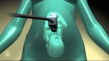
Ultrasound Anatomy Scan images have actually shown to be a great technique to assist minimally intrusive treatments. They have actually ended up being an extremely helpful diagnostic instrument in Obstetrics.
Ultrasound scanner images are utilized to analyze the body. The purpose of the analysis is to discover issues with muscles, tendons, ligaments, joints and soft tissue.
Ultrasound Anatomy Scan will frequently show motion, anatomy and function. It will permit radiologists to evaluate a multiplicity of conditions, and evaluate damage after an injury or disease.
Rare Ectopic Pregnancy Diagnosis During the Ultrasound Anatomy Scan
You may require to have treatment straightaway if an ultrasound scan programs that you have an ectopic pregnancy.
An ectopic pregnancy is a lethal condition where the fertilized egg is beyond your womb so can’t establish effectively. See our different subject on ectopic pregnancy to read more about it.
According to signs you explain, your midwife or physician will typically diagnose, if you have an ectopic pregnancy, when you’re 6 to 10 weeks pregnant.
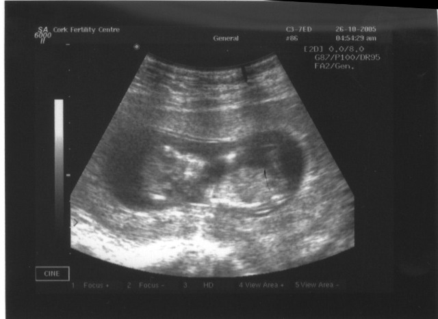
The 12-13 Week Screening Pregnancy Ultrasound
At 12-13 weeks the majority of the primary structures of the child have actually formed. This permits a preliminary physiological study to be carried out. At this phase, numerous gross physiological issues can be found by early comprehensive pregnancy ultrasound scanning.
Case in points are acrania (when the upper part of the child’s skull and brain have actually not established). Also, cases seen are omphalocele and gastroschisis (when the anterior stomach wall has actually not established appropriately).
Pregnancy ultrasound scanning assists in identifying any serious issues with their child’s health in the very first trimester. By doing this, the Moms and Dads are given the chance, extremely early in the pregnancy, to understand the implications of any issues with fetus development. This will allow them to choose how they want to continue with their pregnancy and if necessary look for suitable care.
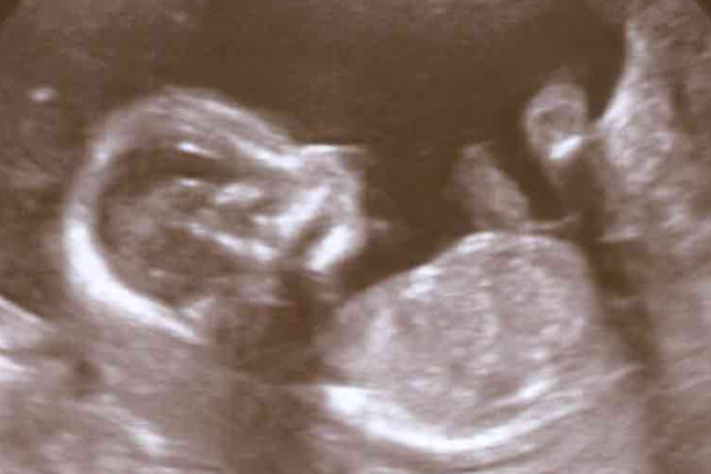
The 18-20 Week Screening Pregnancy Ultrasound
The 18-20 week screening pregnancy ultrasound is a screening test to analyze the advancement of the fetus.
The fetus has regular advancement in 98-99% of pregnancies.
A small number of 18-20 week screening pregnancy ultrasound anatomy scans (1-2%) will recognize a significant structural problem.
Ultrasound Anatomy Scanners
Ultrasound Anatomy Scanners include a console consisting of
- a computer system and electronic devices,
- a video display screen, and
- a transducer that is utilized to scan the body.
The transducer is a little hand-held gadget that looks like a microphone, connected to the scanner by a cable.
On the huge bulk of events, ultrasound anatomy scans work, non-invasive and can assure moms and dads about the health of their coming kid.
They are precise at dating the conception and birth, and offer medics and Moms and Dads the chance to get a look at any significant physical irregularities.
They likewise permit Moms and Dads to see their coming kid in the womb.
Ultrasound Waves
The capacity for ultrasonic imaging of things, with a 3 GHZ acoustic wave producing resolution similar to an optical image, was acknowledged by Sokolov in 1939. However, methods of the time produced fairly low-contrast images with bad level of sensitivity.
Ultrasonic imaging utilizes frequencies of 2 megahertz and greater. The much shorter wavelength permits resolution of tiny internal information in tissues and structures.
The power density is usually less than 1 watt per square centimetre. This prevents heating and cavitation impacts in the things under examination.
High and ultra high ultrasound waves are utilized in acoustic microscopy, with frequencies approximately 4 ghz.
When ultrasound waves are sent out into the body a few of them get better when they struck tissues of varying density.
The time it takes the shown ultrasound waves to go back to the device is equated into a picture of the internal organ, or of the fetus. This image is called an sonogram.
Ultrasound Imaging
Ultrasound Anatomy Scan imaging is based upon the exact same concepts associated with the sounding devices utilized by anglers, bats and ships. When an acoustic wave strikes a things, it bounces backwards, or echoes.
By determining these echo waves it is possible to identify how far away a thing is. And, its size, shape, consistency (whether the item is strong, filled with fluid, or both) and harmony.
With existing innovation advancements, brand-new possibilities in ultrasound imaging are occurring that might enhance diagnostics. In fact experts insist that ultrasound scanning can only be properly carried out. And, its medical efficiency can only ever be enhanced. When users have an extensive understanding of the underlying technical equipment.
Various Types of Ultrasound
The ultrasound that we have actually explained up until now provides a 2 dimensional image, or “piece,” of a 3 dimensional item (fetus, organ). Two other kinds of ultrasound are presently in use.
These are;
- 3D ultrasound imaging, and
- Doppler ultrasound.
Conclusion
Ultrasound Anatomy Scan is pain-free and safe. It also produces images of the baby within the body, utilizing acoustic waves.
Ultrasound imaging, likewise called ultrasound scanning or sonography, includes using a little transducer (probe) and ultrasound gel positioned straight on the skin.
High-frequency acoustic waves are transferred from the probe through the gel into the body. The transducer gathers the noises that get returned.
A computer system then utilizes those acoustic waves to produce an image. But, ultrasound anatomy scan assessments do not utilize ionizing radiation (as utilized in x-rays). Hence, there is no radiation exposure to the mother or child.

Oh! Joy. This takes me back to that very first scan. Now, I reminisce. We have 3 boys and a girl. All grown up now of course.
Trish